Angle-Resolved Photoemission Spectroscopy
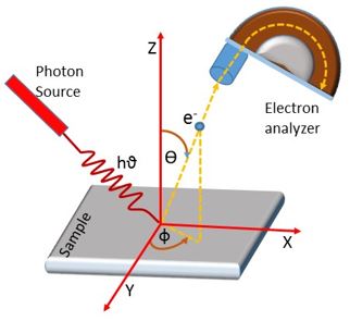
Angle-resolved photoemission spectroscopy (ARPES) is a powerful spectroscopic technique based on Einstein’s photoelectric effect (“photon in, electron out”), first observed by Hertz in 1887. When incident photons have sufficient energy to overcome the binding energy of electrons in a material, they can eject photoelectrons from the surface. By measuring the kinetic energy and the polar and azimuthal emission angles of these electrons at a fixed photon energy, one can determine their binding energy and in-plane momentum. This enables precise reconstruction of the material’s electronic structure.
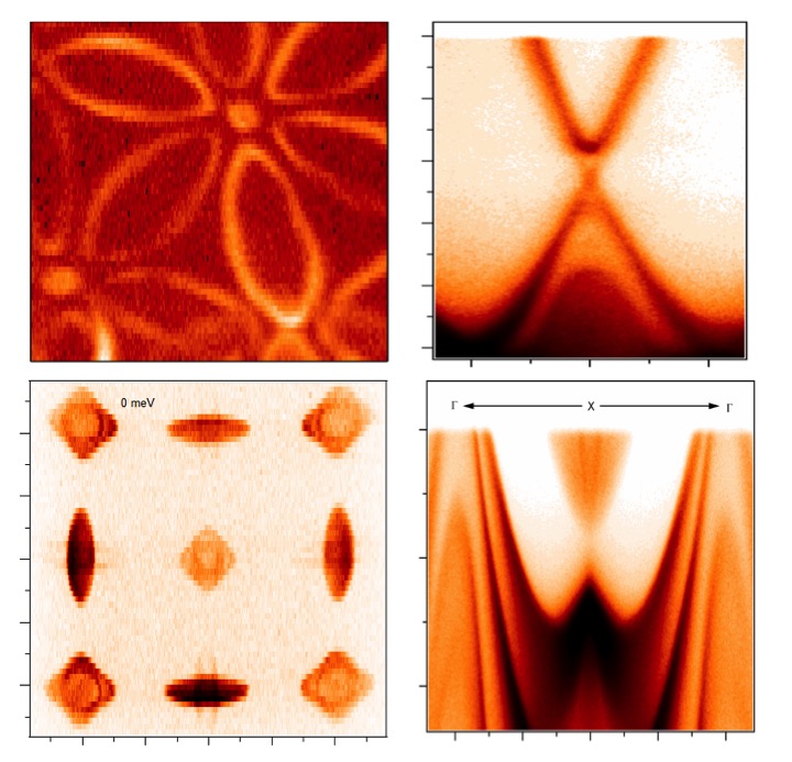
ARPES measures the occupied states of the single-particle spectral function, providing detailed insight into band dispersions and many-body interactions. It can map band dispersion, determine band gaps, and image Fermi surfaces. Spin-resolved ARPES (Spin-ARPES) extends this capability by revealing the spin polarization of electronic states. Because of its extreme surface sensitivity, ARPES has become a key tool for studying topological materials. For example, the robust surface states of topological insulators—characterized by spin–momentum–locked Dirac cones—can be directly visualized using ARPES.
ARPES systems can be driven by a variety of light sources, including synchrotrons, lasers, and helium discharge lamps. Our group frequently conducts systematic spectroscopic measurements at synchrotron facilities such as the Advanced Light Source (ALS) at Berkeley, the Stanford Synchrotron Radiation Lightsource (SSRL) at Stanford, and the Swiss Light Source (SLS) in Switzerland. In addition, our home-built ARPES system is equipped with a helium discharge lamp and a high-harmonic–generation laser source, enabling complementary laboratory-based measurements.
Pump–Probe Setup
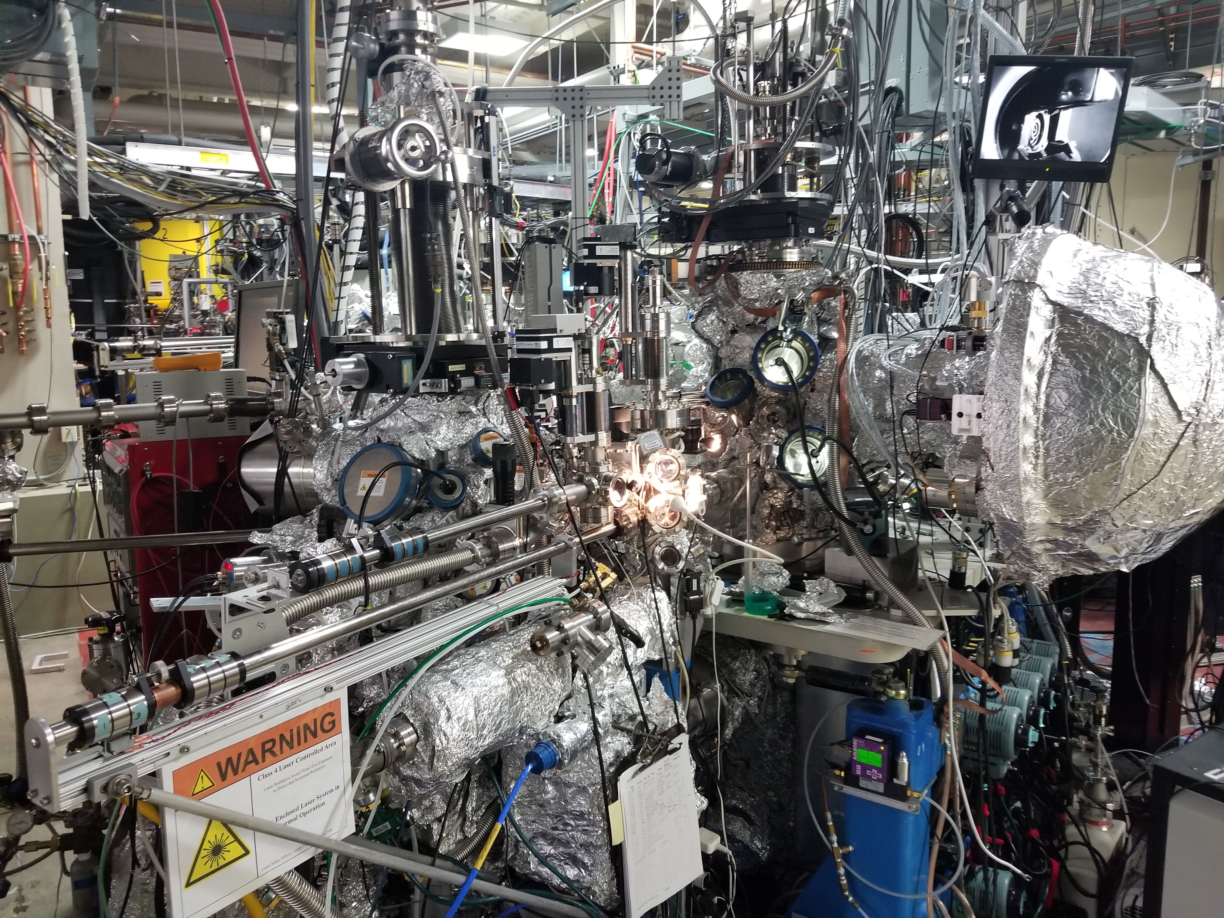
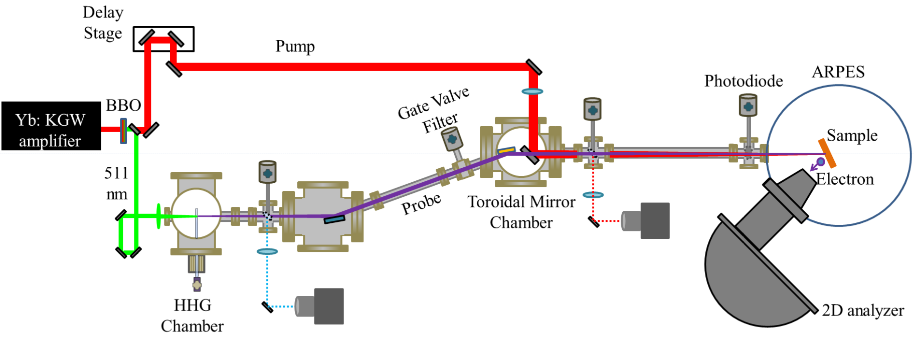
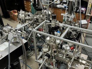
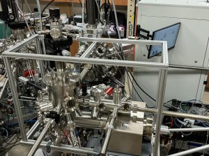
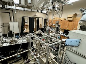
Time- and angle-resolved photoemission spectroscopy (tr-ARPES) is a powerful technique for studying quantum materials, as it can probe nonequilibrium states and track ultrafast dynamics on femtosecond to attosecond timescales. In collaboration with Professor Chini, we have developed a novel high-order-harmonic–based tr-ARPES system, whose layout is shown below.
Second-harmonic pulses are generated using a 2 mm–thick BBO crystal with more than 50% conversion efficiency and are focused by an f = 175 mm lens into a krypton-filled gas cell (0.4 mm in length) at a backing pressure of 20 torr to produce high harmonics. For trARPES measurements, isolation of a single harmonic is essential. In our setup, this is achieved by blocking lower harmonic orders with a 500 nm–thick Al foil filter and suppressing the high-harmonic cutoff by reducing the laser intensity.
The tunable repetition rate of the laser enables a reduction in pulse energy while increasing the repetition rate—maintaining constant average power. This allows the 9th harmonic to be cleanly isolated, achieving >10× extinction of neighboring 7th and 11th harmonics, with a repetition rate tunable between 75 and 120 kHz. The harmonic probe is directed by a SiC-coated flat mirror and focused onto the sample using an f = 650 mm toroidal mirror. The on-target photon flux of the 9th harmonic is measured to be approximately 5 × 10¹⁰ photons/s.
The residual 1030 nm laser beam after the BBO crystal serves as the pump pulse. The pump–probe delay is controlled via a motorized delay stage in the pump arm, enabling precise temporal resolution for ultrafast measurements.
Angle- and time-resolved ARPES
Single Crystal and Thin Film Growth
We employ various techniques to grow high-quality single crystals of topological materials. For thin films of quantum materials, we use molecular beam epitaxy (MBE)—a method that is directly integrated with our ultrafast pump–probe spectroscopy setup.
Our research focuses on emergent quantum materials, particularly their light–matter interactions, to explore the underlying electron dynamics. To achieve this, we have established a state-of-the-art MBE system that connects directly to our spectroscopic equipment, enabling in situ growth and characterization.
Molecular beam epitaxy is a sophisticated crystal growth technique capable of producing ultrahigh-quality thin films under ultrahigh vacuum (UHV) conditions, with precise control over thickness, composition, and morphology. MBE is widely recognized as one of the best techniques for growing crystalline films for advanced materials research.
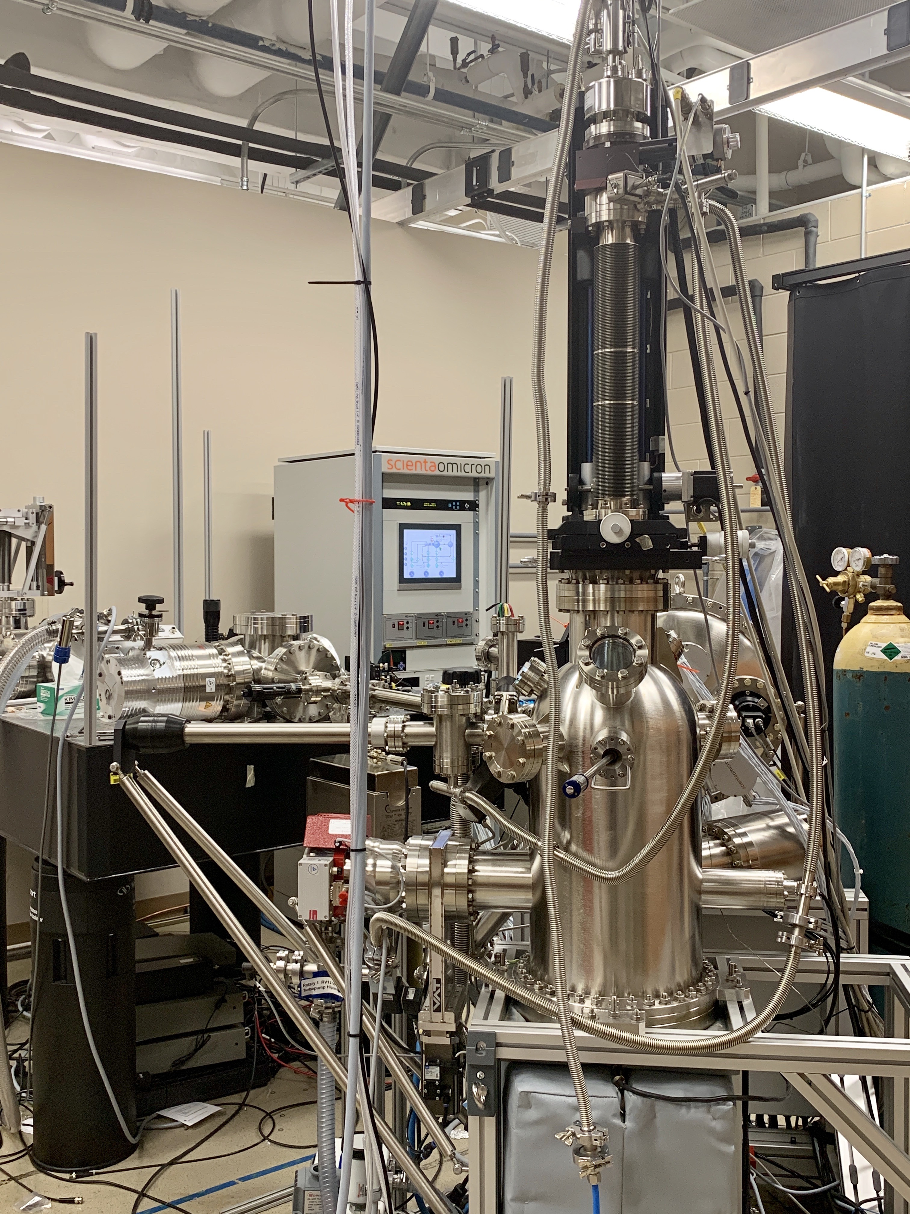
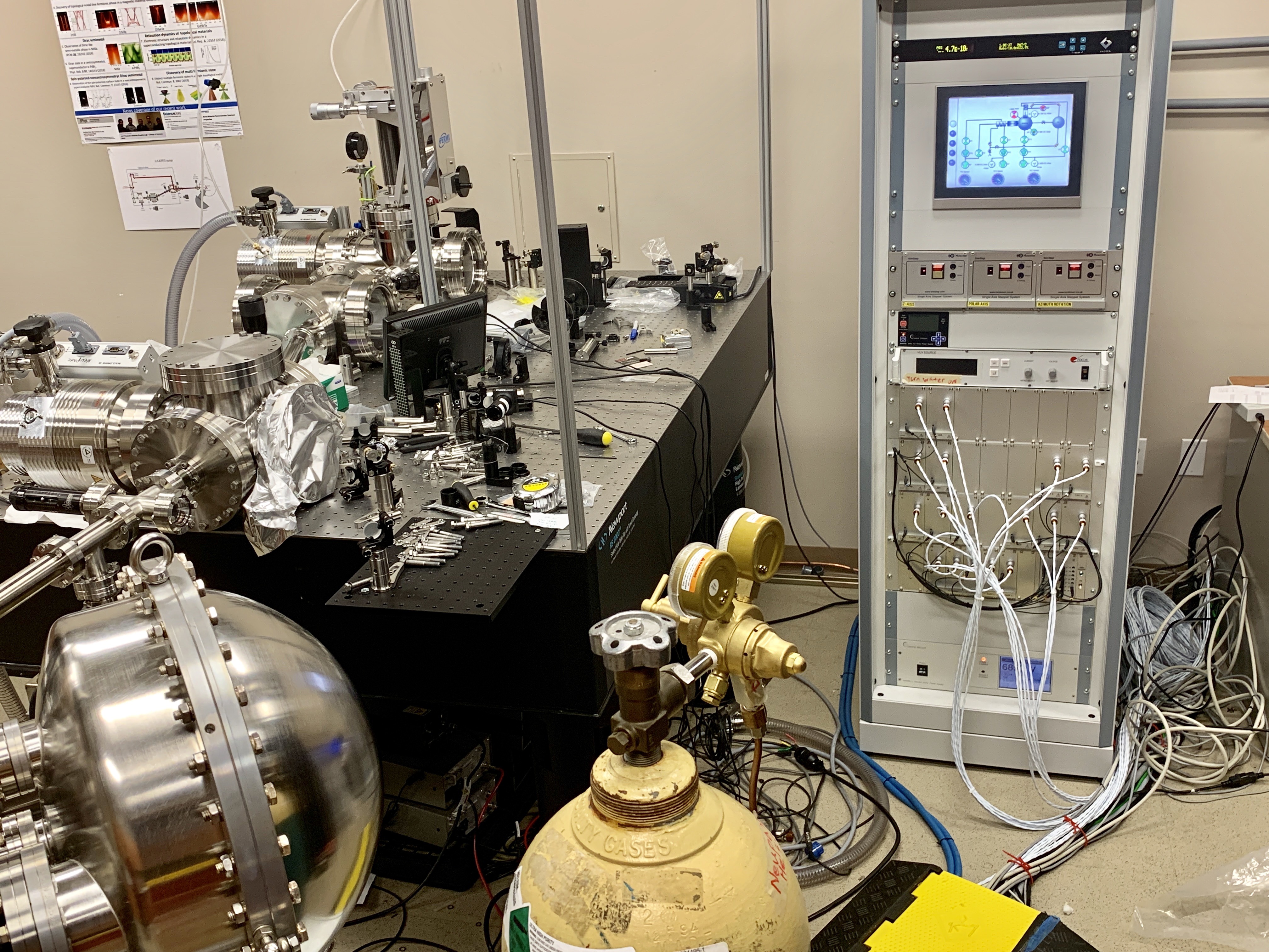
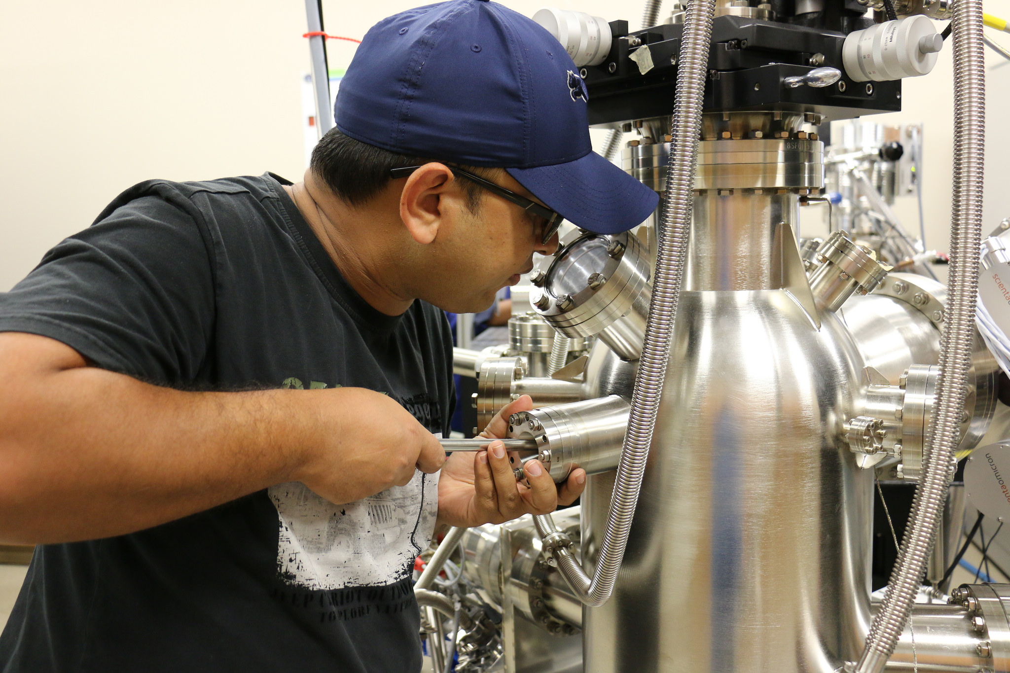
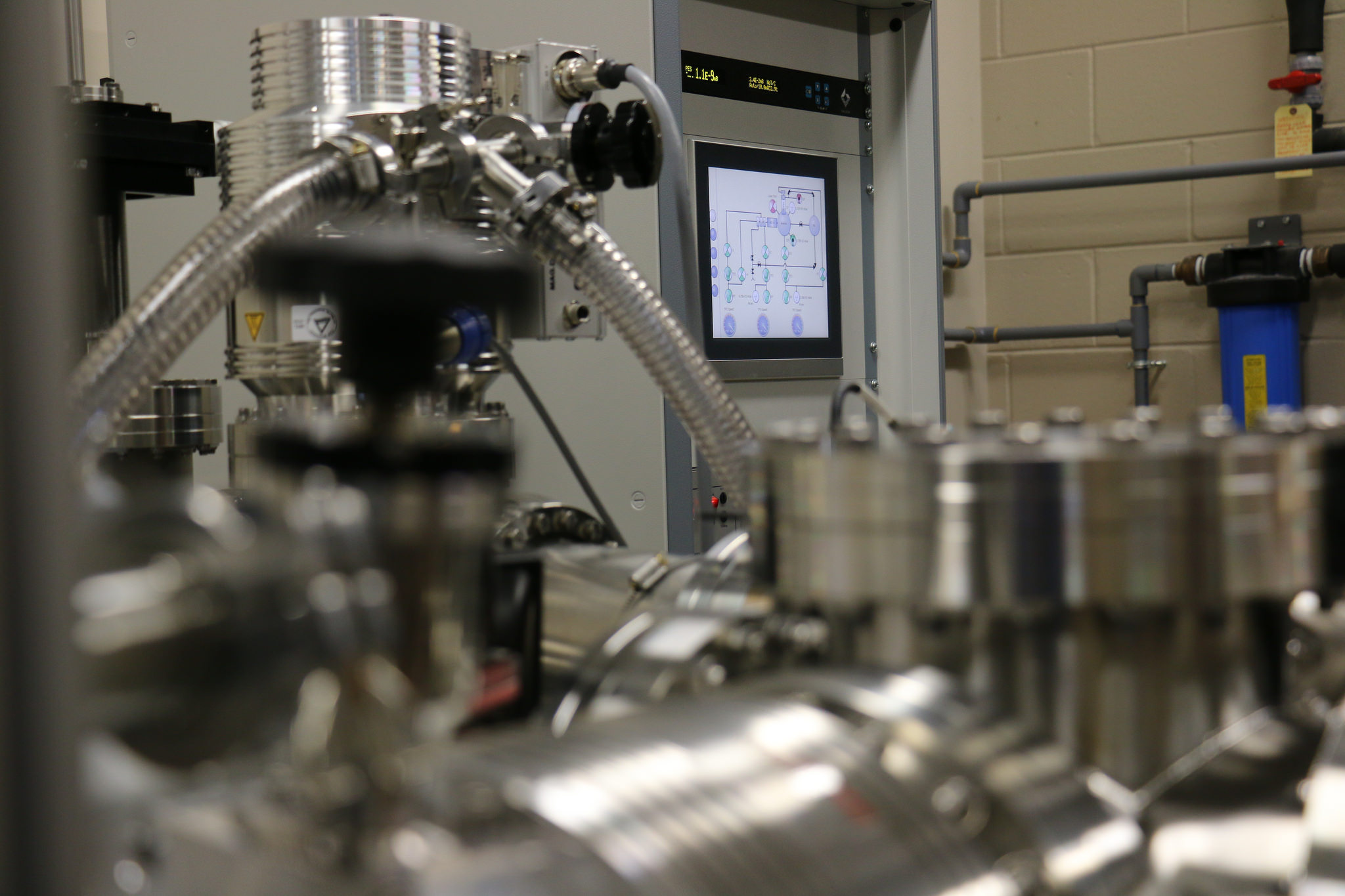
MBE Equipment @ Neupane Lab
We use the LAB10 Molecular Beam Epitaxy System from Scienta Omicron—a small-sample deposition tool designed for exploratory material research under UHV conditions. The system supports sample sizes up to 10 × 10 mm and is highly versatile, accommodating a wide range of MBE applications, including the deposition of metals, two-dimensional materials, and topological quantum materials. The LAB10 system is fully integrated with our ultrafast spectroscopy chamber, enabling seamless studies of thin films from growth to ultrafast optical and photoemission measurements.

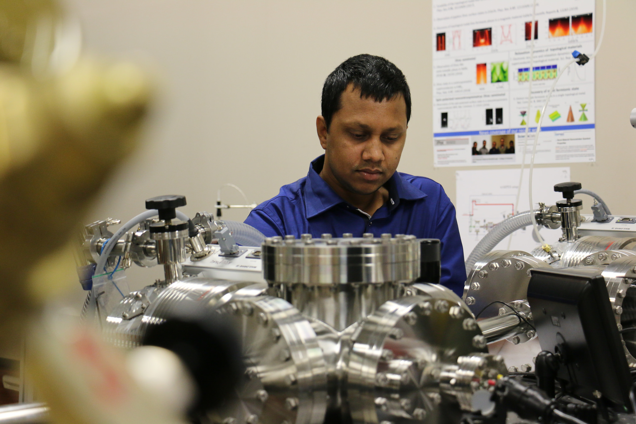
Beamlines
Our group performs measurements at various beam lines at ALS, SSRL, NSLS and SLS for advanced spectroscopic characterization of topological quantum materials.