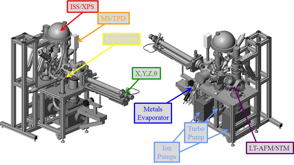
UHV surface-science chamber housed in the Kaden Lab. Photo Credit to Joshua Sweet.
In Situ Techniques:
Scanning-Tunneling Microscopy/Spectroscopy: Provides atomic-scale structural and near-Fermi electronic characterization of near-surface features by monitoring tip height fluctuations needed to maintain constant electron tunneling currents while rastering the tip parallel to the surface or changes in tunneling current while varying the tip-sample bias at a fixed position. The miracle of this technique lies in the development of piezo-electric materials allowing for the controllable and reproducible movement of the tip at sub-angstrom length-scales.
Atomic-Force Microscopy/Spectroscopy: Provides complementary structural insights by monitoring fluctuations in long-range attractive forces between surface atoms and a resonantly oscillating probe. Related techniques include Kelvin Probe and Single Electron Tunneling Electrostatic Force Spectroscopy, which provide local measures of fluctuations in work-function and threshold valence/conduction energies for local states isolated near the surface of insulating materials. Since these techniques utilize fluctuations in tip-sample forces rather than current-based feedback, they are functional of a wider range of materials than STM/S, which require electrically conductive samples.
X-ray Photoemission Spectroscopy: Provides a long-range methodology for characterizing the elemental composition and charge-state of atoms residing in the near-surface region of samples based on the quantized nature of the photoelectric effect. In short, a fixed energy photon excitation source is directed towards the sample, resulting in several processes, among them core-electron ejection. The kinetic energy of these electrons is then related to their atom-specific core-level binding energies, and more subtle shifts in these values can be further related to changes in hybridization, charge-state, and the degree of relaxation after creating the core-hole. In addition to core-levels, the technique also allows for investigation of valence-shell electrons in the near-Fermi region (albeit with larger background contributions relative to other techniques, like UPS), secondary Auger electrons created via one of many core-hole relaxation processes, and work-function determination via analysis of the low kinetic energy threshold for inelastically scattered secondary electrons. Additional structural effects may be elucidated via peak intensity comparisons as a function of variable photon energy or (more appropriate to our group) electron collection angle. Finally, a more thorough understanding of the initial-state contributions (the effects causing changes to the electronic structure of atoms in the sample prior to photo-emission) to the observed core-level shifts may be revealed via an appropriate application of an Auger parameter analysis.
Ion Scattering Spectroscopy: Provides a measure of the relative atomic concentration profile of the atomic surface layer of a sample by probing the kinetic energy lost to recoil after colliding low energy ions (typically He+ at 1 keV) from various atomic nuclei present at the interface. The technique essentially provides a vertical projection of the two-dimensional surface footprint occupied by various atomic constituents and derives its extremely high degree of surface sensitivity from the exceedingly small survival probability of He cations following nuclear collisions with other atoms.
Mass Spectroscopy/Temperature Programmed Desorption: Provides the relative gas-phase concentration of molecules near the sample surface as a function of mass by first ionizing neutral species via electron impact and then electrostatically guiding the resultant products into a quadruploe mass filter, where they are seperated on the basis of variation in mass-to-charge ratio. This allows us to monitor vacuum conditions, and probe various gas-phase products of interest evolved during catalytically relevant reactions.
Low Energy Electron Diffraction: Provides the long-range repeating atomic surface-structure of planar crystalline interfaces by irradiating a small (millimeter order of magnitude) area of the sample with a collimated beam of electrons, which then reflectively diffract in specific angular directions dependent on the lateral unit cell dimensions and beam energy, and can be detected using a phosphor screen.

